Human Leg Muscles Diagram - Anatomy Leg Muscles Diagram Quizlet / This diagram depicts anatomy of the leg muscles.human anatomy diagrams show internal organs, cells, systems, conditions, symptoms and sickness information and/or tips for healthy living.
Human Leg Muscles Diagram - Anatomy Leg Muscles Diagram Quizlet / This diagram depicts anatomy of the leg muscles.human anatomy diagrams show internal organs, cells, systems, conditions, symptoms and sickness information and/or tips for healthy living.. Muscle and bone anatomy 12 photos of the muscle and bone anatomy back muscles and bones anatomy, human muscle and bone anatomy, muscle & bone anatomy 3d free download, muscle and bone anatomy app, muscle and bone anatomy quiz, human muscles, back muscles and bones anatomy, human muscle and bone anatomy, muscle & bone. They allow you to move and provide support for your upper body. The thigh (proximal lower limb) muscles are arranged into three compartments : The calf muscle, on the back of the lower leg, is actually made up of two muscles: See more ideas about muscle anatomy, human anatomy and physiology, body anatomy.
Human anatomy diagrams show internal organs, cells, systems, conditions, symptoms and sickness information and/or tips for healthy living. The hamstring muscle attachment points. The below the gluteus medius are several muscles, one of which is the gluteus minimus, the smallest of the gluteal muscles. Leg muscles anatomy leg anatomy human body anatomy human anatomy and physiology anatomy study anatomy reference thigh muscles body muscle anatomy muscular system anatomy. It is a synergist for the gluteus medius.
3 from Medial compartment, also known as adductor compartment; Your leg muscles are some of the hardest working muscles in your body. The hamstring muscle attachment points. The knee is one of the largest and most complex joints in the body. The quad muscles— which form the meaty mass on the front of your thighs — are among your strongest muscle groups, and play a critical role in athletic activities. The calf muscle, on the back of the lower leg, is actually made up of two muscles: Climbing stairs, standing, walking, and running are all activities that require strong contractions from the posterior muscle group to extend the leg. Human anatomy diagrams show internal organs, cells, systems, conditions, symptoms and sickness information and/or tips for healthy living.
The gastrocnemius is the larger calf muscle, forming the bulge visible beneath the skin.
It is controlled by the obturator nerve. One of the most important tendons in terms of mobility of the leg is the achilles tendon. The muscles of the human body can be categorized into a number of groups which include muscles relating to the head and neck, muscles of the torso or trunk, muscles of the upper limbs, and muscles of the lower limbs. The thigh (proximal lower limb) muscles are arranged into three compartments : Hamstrings (made of 3 muscles): The four muscles that make up the quadriceps are the strongest and leanest of all muscles in the body.these muscles at the front of the thigh are the major extensors (help to extend the leg. Your legs are two of your most important body parts. The knee is one of the largest and most complex joints in the body. The human leg, in the general word sense, is the entire lower limb of the human body, including the foot, thigh and even the hip or gluteal region. The gluteus medius muscle helps abducts the thigh along with the gluteus maximus, but can rotate the thigh inward where the gluteus maximus rotates the thigh outward. The gastrocnemius is the larger calf muscle, forming the bulge visible beneath the skin. Raises heal when leg is straight. Muscle of the human leg diagram in this image, you will find muscle of the human leg diagram, hip and femur middle layer, hip and femur deep layer, overview of the most important muscles of the leg, femur middle layer, femur deep layer, rectus femoris m.
This diagram depicts anatomy of leg muscles. It is controlled by the obturator nerve. Muscle of the human leg diagram in this image, you will find muscle of the human leg diagram, hip and femur middle layer, hip and femur deep layer, overview of the most important muscles of the leg, femur middle layer, femur deep layer, rectus femoris m. Raises heal when leg is straight. This diagram depicts anatomy of the leg muscles.human anatomy diagrams show internal organs, cells, systems, conditions, symptoms and sickness information and/or tips for healthy living.
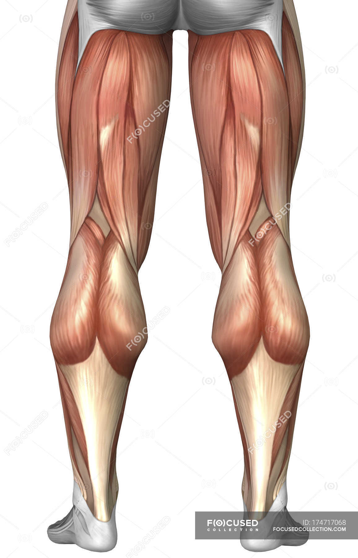
Diagram Illustrating Muscle Groups On Back Of Human Legs Vastus Lateralis Gracilis Stock Photo 174717068 from st.focusedcollection.com It is controlled by the obturator nerve. Muscle anatomy in foot 12 photos of the muscle anatomy in foot muscle anatomy human foot. The quad muscles— which form the meaty mass on the front of your thighs — are among your strongest muscle groups, and play a critical role in athletic activities. Anterior compartment, also known as the extensor compartment; Just need a glimpse, leave your valuable advice let us know , and subscribe us! The muscles of the lower leg can divided into 3 main groups: These four muscles at the front of the thigh are the major extensors (help to extend the leg. In the leg muscles diagram above, there are many muscles that make up your legs and support it to move.
Legs are used for standing, and all forms of.
Biceps femoris (long head) biceps femoris (short head) semitendinosus. This diagram depicts anatomy of leg muscles. The following diagram illustrates the actions of the terms adduction, abduction, flexion and extension at the different joints. Anterior muscles of the lower leg, lateral fibularis group and posterior muscles of the lower le. Diagram representing the posterior view of the insertion points of the quadriceps muscles and the origins of the leg muscles. These four muscles at the front of the thigh are the major extensors (help to extend the leg. The human leg, in the general word sense, is the entire lower limb of the human body, including the foot, thigh and even the hip or gluteal region. The human body has three different types of muscles. Muscle charts of the human body for your reference value these charts show the major superficial and deep muscles of the human body. The calf muscle, on the back of the lower leg, is actually made up of two muscles: Leg muscles anatomy leg anatomy human body anatomy human anatomy and physiology anatomy study anatomy reference thigh muscles body muscle anatomy muscular system anatomy. The hamstring muscle attachment points. It is controlled by the obturator nerve.
The gluteus medius muscle helps abducts the thigh along with the gluteus maximus, but can rotate the thigh inward where the gluteus maximus rotates the thigh outward. Hamstrings (made of 3 muscles): It is a synergist for the gluteus medius. Leg muscles anatomy leg anatomy human body anatomy human anatomy and physiology anatomy study anatomy reference thigh muscles body muscle anatomy muscular system anatomy. Anterior compartment, also known as the extensor compartment;
Illustration Of The Human Thigh Muscles Download Scientific Diagram from www.researchgate.net Human anatomy diagrams show internal organs, cells, systems, conditions, symptoms and sickness information and/or tips for healthy living. Tutorials and quizzes on muscles that act on the leg/ leg muscles (tibia & fibula), using interactive animations and labeled diagrams. We are pleased to provide you with the picture named anatomy of the groin area superficial muscles and deep muscles.we hope this picture anatomy of the groin area superficial muscles and deep muscles can help you study and research. The muscles of the lower leg can divided into 3 main groups: One travels down each leg and. Muscle anatomy in foot 12 photos of the muscle anatomy in foot muscle anatomy human foot. One of the most important tendons in terms of mobility of the leg is the achilles tendon. Muscle and bone anatomy 12 photos of the muscle and bone anatomy back muscles and bones anatomy, human muscle and bone anatomy, muscle & bone anatomy 3d free download, muscle and bone anatomy app, muscle and bone anatomy quiz, human muscles, back muscles and bones anatomy, human muscle and bone anatomy, muscle & bone.
Posterior compartment, also known as the flexor compartment;
In the leg muscles diagram above, there are many muscles that make up your legs and support it to move. The four muscles that make up the quadriceps are the strongest and leanest of all muscles in the body.these muscles at the front of the thigh are the major extensors (help to extend the leg. This anatomy chart is a great example of beauty and function in one, as it is pleasing to look… Biceps femoris (long head) biceps femoris (short head) semitendinosus. Just need a glimpse, leave your valuable advice let us know , and subscribe us! Included are several layered views of the back muscles, the dorsal muscles, subclavius muscles, rhomboideus major and minor muscles, deltoid muscles and many more. One of the most important tendons in terms of mobility of the leg is the achilles tendon. Posted on may 22, 2016 by admin. It is a synergist for the gluteus medius. Climbing stairs, standing, walking, and running are all activities that require strong contractions from the posterior muscle group to extend the leg. Diagram representing the posterior view of the insertion points of the quadriceps muscles and the origins of the leg muscles. The knee joins the thigh bone (femur) to the shin bone (tibia). This important tendon in the back of the calf and ankle stores the elastic energy needed for running, jumping, and other physical activity.
See more ideas about muscle anatomy, human anatomy and physiology, body anatomy human muscles diagram. Included are several layered views of the back muscles, the dorsal muscles, subclavius muscles, rhomboideus major and minor muscles, deltoid muscles and many more.


0 Comments